
Editor'S Choice
 resistance
Krperprozesse Clarithromycin
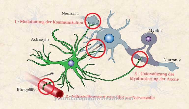
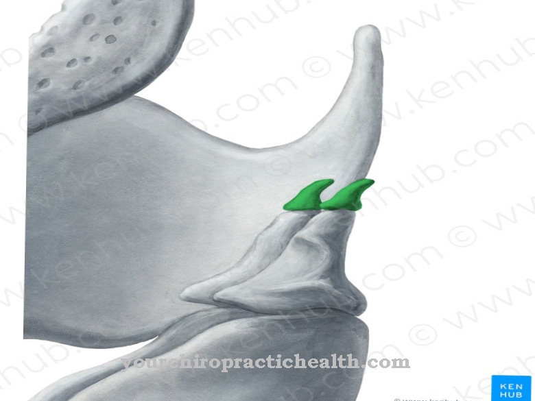
Active Ingredients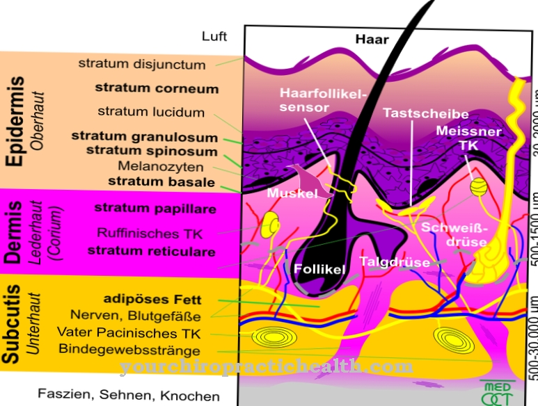 Meissner corpuscles
Anatomy Adonis
Medicinal Plants Living in constant worry: When fear dominates everyday life
Counselor
Most Visited
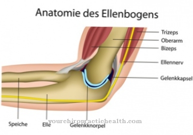 Broken elbow
Diseases Ciguatera fish poisoning
DiseasesLangerhans Islands
Anatomy transcription
Krperprozesse Cryopyrin-Associated Periodic Syndrome
Diseases
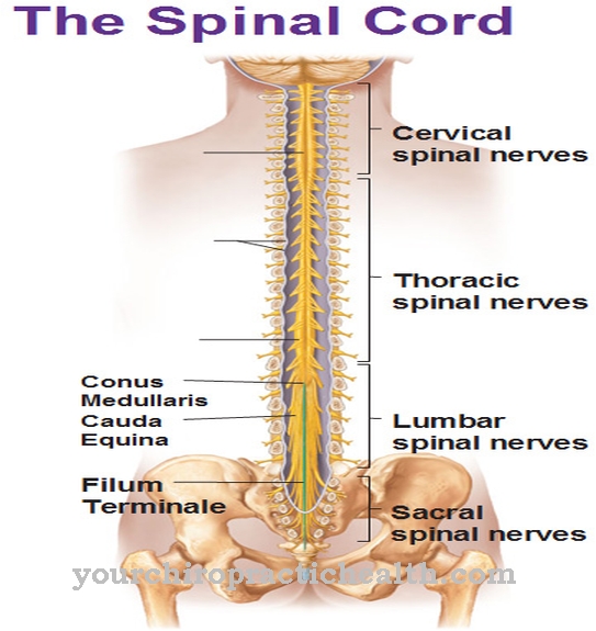
The conus medullaris is the conical end of the spinal cord. Paraplegia at the medullary conus is known as cone syndrome and results in various disorders that are due to the failure of the spinal cord nerves supplying it. The illness
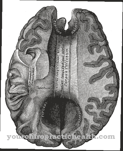
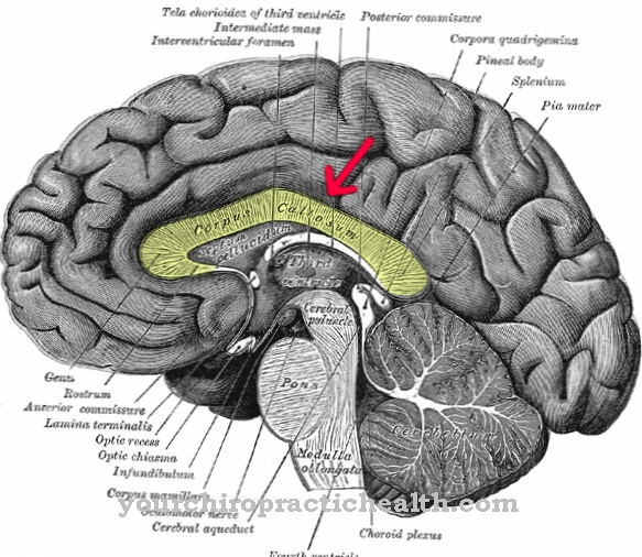
The corpus callosum connects the hemispheres of the brain. It runs transversely and consists of a multitude of fibers. It is also called a beam.
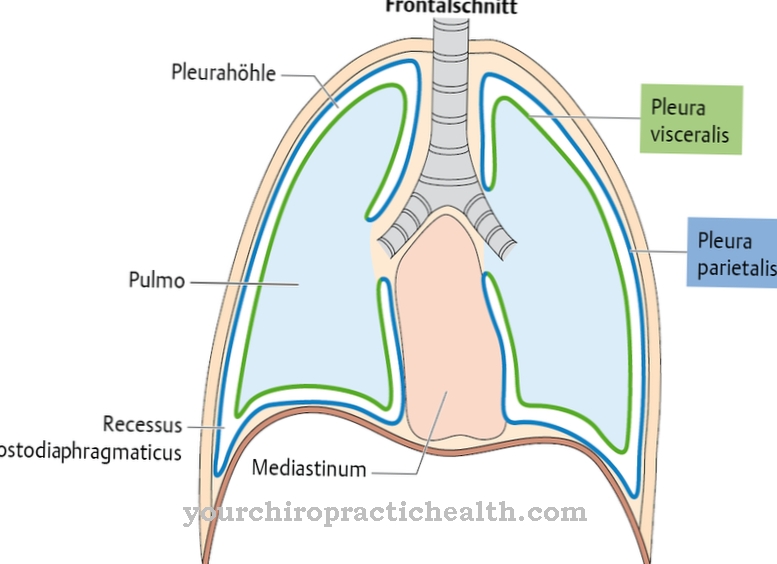
The gap between the inner and outer sheets of the pleura (pleura) is called the pleural cavity. The pleural cavity is filled with fluid so that the two pleural leaves do not rub against each other. With excessive fluid accumulation in

The corpus ciliare is also known as the ciliary body or ray body and is located in the middle skin of the eye. It is used for accommodation, aqueous humor production and lens suspension. If the hanging fibers of the lens break in an accident, can
The corpus mamillare is a structure in the interbrain (diencephalon) and forms part of the limbic system. It is also the origin of the mamillothalamic and mamillotegmental tracts. Damage to the corpus mamillare can
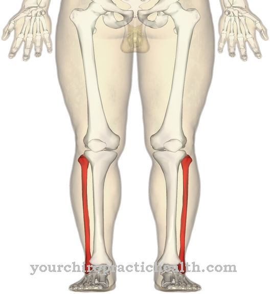
One of the two lower leg bones is called the fibula. This belongs to the long bones.
Long bones get their name because of their elongated shape. The bones have a uniform marrow cavity in which the bone marrow is located. They only occur in the extremities.
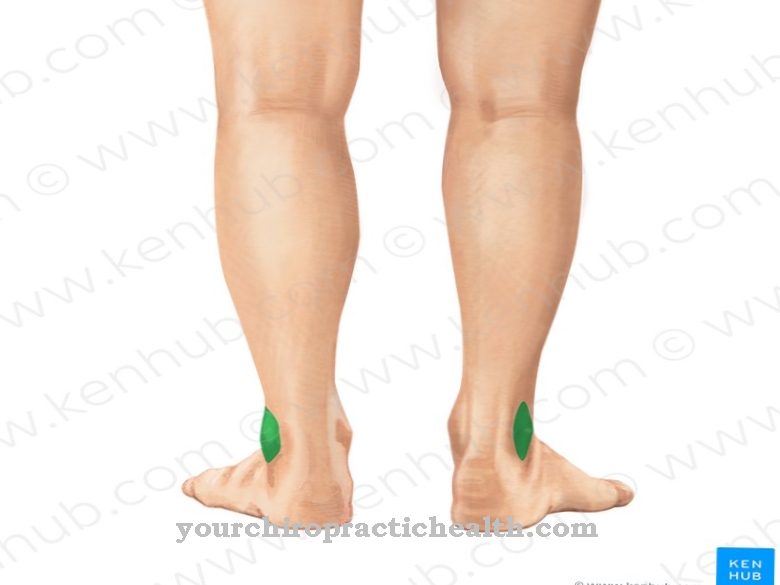
The lateral malleolus is the thickened end of the fibula that is involved in the ankle joint. This so-called outer ankle creates the conditions for the flexion and extension of the foot in a dorsal and plantar direction. Fractures of the ankle joint
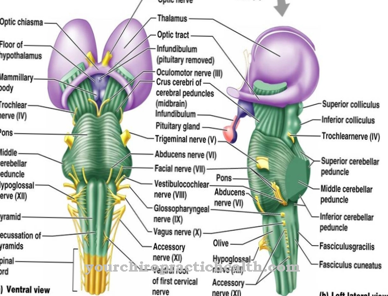
The crura cerebri form the two cerebral legs and represent part of the midbrain. They contain fibers of the internal capsule, through which nerve tracts from different areas of the brain mainly lead to the bridge (pons). Damage to these nerve fibers
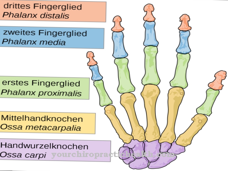
The finger bones are among the bony structures of the upper extremities of the human musculoskeletal system. With the exception of the thumb, all fingers consist of three individual bones (phalanges) that are connected by joints.
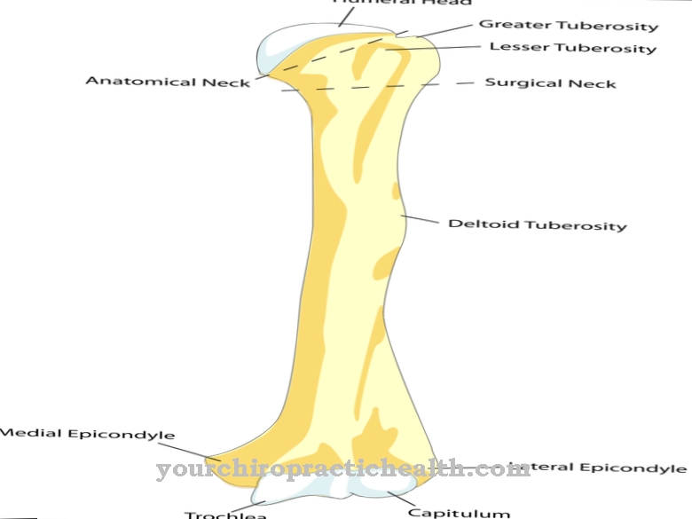
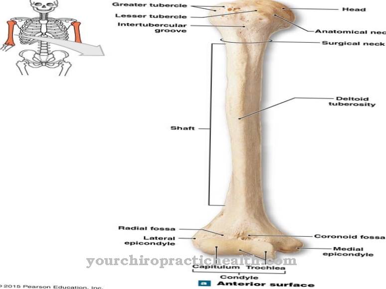
The humerus is the upper arm bone, one of the strongest bones in the upper extremities. Nerves and blood vessels run along the humerus and numerous muscles have their sinewy attachment here. Despite its enormous stability, fractures in the humerus are not uncommon
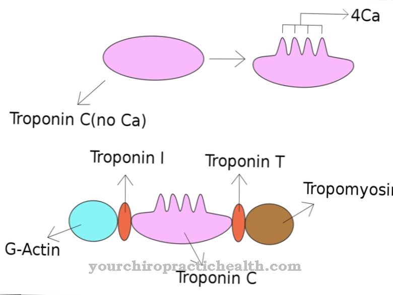
Troponin is a complex of three globular protein subunits. As part of the contractile apparatus of the muscle, troponin regulates muscle contraction. It is of particular importance in the diagnosis of heart attacks.
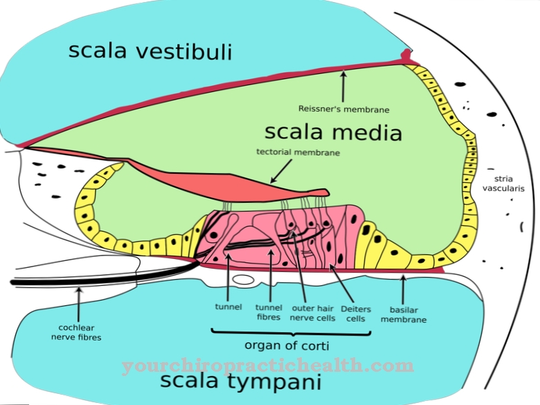
The organ of Corti is located in the inner ear in the cochlea and consists of supporting cells and sensory cells that are responsible for hearing. When a sound wave stimulates the hair cells, they trigger an electrical signal in the downstream neuron
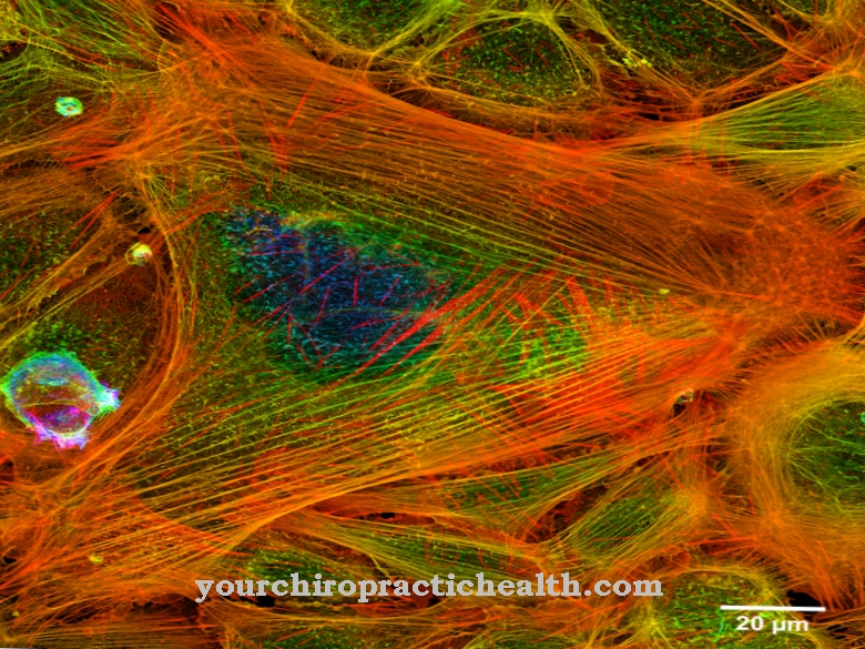
The cytoskeleton consists of a dynamically changeable network of three different protein filaments in the cytoplasm of the cells.
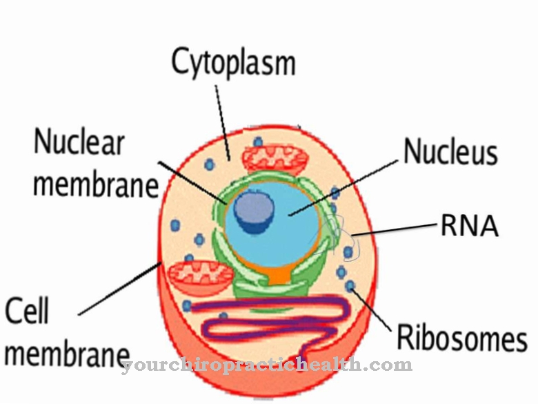
The cytoplasm fills the interior of a human cell. It consists of the cytosol, a liquid or gel-like substance, the organelles (mitochondria, Golgi apparatus, etc.) and the cell skeleton. Overall, the cytoplasm serves the enzymatic
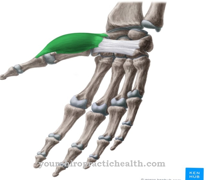
The flexor pollicis brevis muscle is a hand muscle with two heads. He flexes his thumb and participates in his adduction. The striated skeletal muscle receives the nervous signals from the ramus profundis nervi ulinaris and from the nerve

The perineum, or dam, is the area that separates the anus from the genitals. The area is mostly made up of muscles, but has highly sensitive skin. The perineum is therefore also known as the erogenous zone.
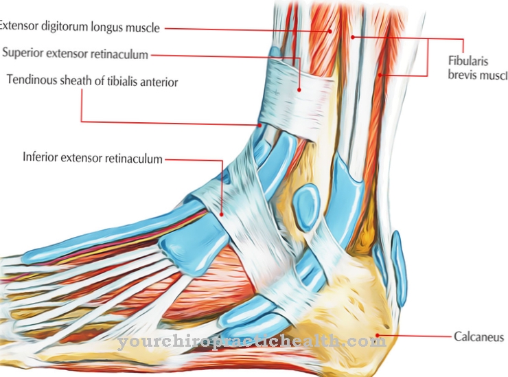
The retinaculum flexorum is a ligament that is composed of relatively firm connective tissue.
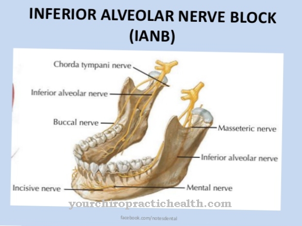
The inferior alveolar nerve is found in the lower jaw and contains sensitive fibers that are responsible for the teeth, chin and lower lip. In addition, the inferior alveolar nerve includes a motor branch, the mylohyoid muscle
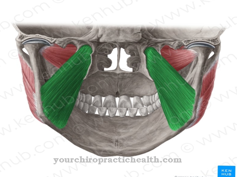
The medial pterygoid muscle is a muscle that belongs to the human chewing muscles. It is located on the inside of the temporomandibular joint. Its function is the movement of the temporomandibular joint.

Interesting Articles
Recommended
Most Popular